
Early Life and Career
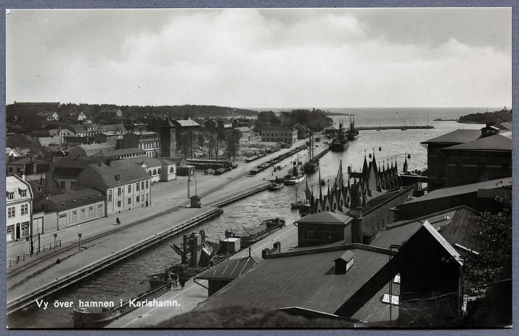
Karlshamn, Sweden, in the 1920s
Per-Ingvar Persson was born in Karlshamn, Sweden, in 1929, to Par Persson and Anna Margit Ingeborg Persson (née Andersson).
He followed his interest in medicine to the University of Lund, pursuing a degree in medical studies in the early 1950s.
He would graduate in 1956 and continued at the university for his PhD.
Throughout his time in medical school, he studied tissue response to injury at the microscopic level as part of his thesis research. In the midst of conducting this research, he discovered that in the lab animals he was studying, he was unable to remove the titanium optical chamber implants he had inserted to his specimen’s leg to more easily view the production of marrow on the microscopic level. The bone had begun to grow around the implant, all on its own!
“One who breaks the ground”
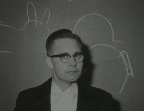
Brånemark giving his dissertation presentation at Lund University, 1959, Image courtesy of the American College of Prosthodontics.
This was an unexpected but fascinating outcome in his research, and in the coming year, this discovery would go on to shape the rest of his career and life.
Per-Ingvar concluded his studies at the university, successfully defending his thesis, titled “Vital Microscopy of Bone Marrow in Rabbit” and officially graduating with his PhD in 1959.
After graduating, Per-Ingvar would go on to change his last name from Persson, to Brånemark, the name he would become known by all over the world.
From Swedish, Brånemark roughly translates to “one who breaks the ground”. This new name would prove to ring true throughout his life and career.
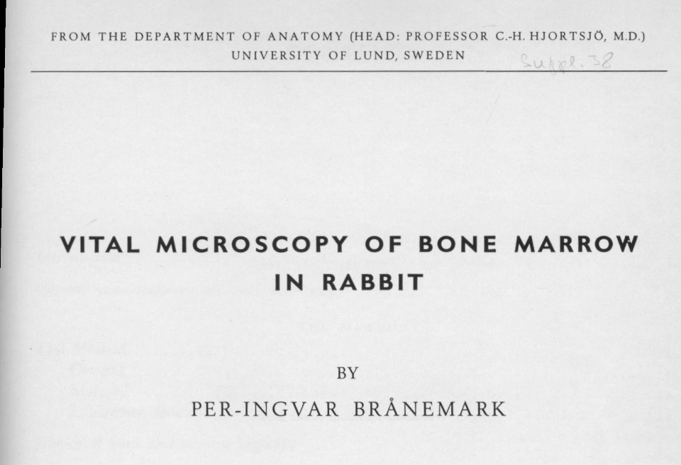
Front page of Per-Ingvar Brånemark’s Thesis titled “Vital Microscopy of Bone Marrow in Rabbit.”
Early Trials in Osseointegration
By 1963, Brånemark started his career at the University of Goteborg, in Goteborg, Sweden, as a Professor of Anatomy. Brånemark spent the 1960s balancing both his teaching duties with research.
His PhD research experience had inspired him, and he continued his research in tissue response to titanium implants, beginning with seeing how bone tissues in the long bones of rabbits would respond to these implants.
From here, he performed trials on canines, investigating how tissue in long bones and even in extremities, like tail bones reacted.
His studies found that bone tissue would consistently grow around these implants, becoming consistently firmly integrated to the point that it became nearly impossible to remove them.
At the time, implants were experimented with on patients orally for tooth loss as a possible alternative to dentures, but sawlittle success. Before Brånemark, implants could be made of metals and synthetic materials like gold, silver, cobalt, chrome, or stainless steel.
But the human body is very finicky and tends to reject foreign materials, like implants, and patients often suffered from rejection from the body, inflammation, and gingivitis. Removing failed implants sometimes caused painful oral trauma. Ultimately, these implants didn’t last their patients long.
Observing this significant gap, Brånemark realized his success in animal trials might mean titanium could be a viable option for successful, long-term oral implants.
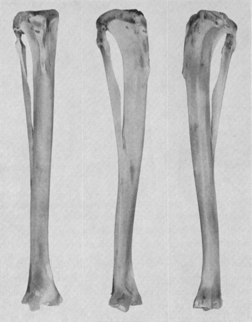
Image from Brånemark’s Thesis, Rabbit Tibiofibula, 1959
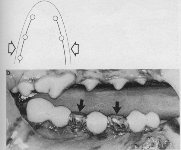
Canine mouth with several implanted fixtures to replace removed teeth, 1969
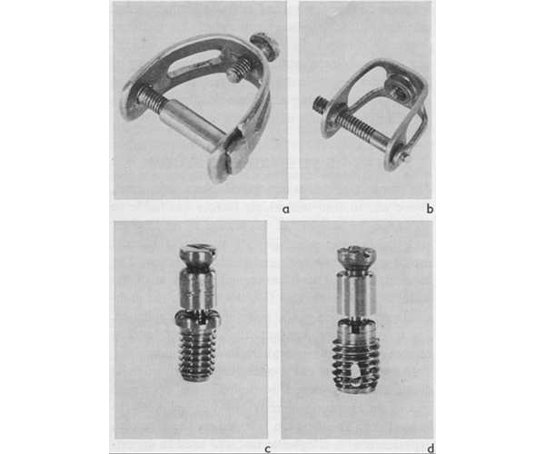
Prototype titanium fixtures created by Brånemark, 1969
He and his team set out to continue their trials, this time using titanium implants orally.
They conducted their work with canines, extracting teeth, allowing complete healing, and implanting specially crafted tooth replacements.
Much like the results he saw previously, these titanium implants healed well, with consistent bone growth around them. Brånemark’s confidence and his hopes were up; his discovery and research might just benefit human patients next.
First Human Patients
The first implant trials on humans were conducted on several volunteers. Brånemark and his team implanted titanium optical chambers into their upper arm bones, which would enable the research team to get a good look at the ways their tissues and cells reacted to the implants.
They saw no signs of rejection. Instead, there was healing and the beginnings of bone growing around these implants. This trial proved a resounding success, and a hopeful excitement spread through Brånemark and his team in this new system’s ability to be used in, and help, human patients. The years of studies, animal trials, and now human experiments were finally paying off.
In 1965, Brånemark worked with his first human patient, a Swedish man named Gosta Larsson. Larsson suffered from lifelong jaw deformities, a cleft palate, and serious tooth problems. He reportedly said on the state of his mouth and his dental problems, he “couldn’t even chew a slice of bread. The few teeth I had were, more or less, randomly distributed through my mouth…”.
Working with Larsson, Brånemark and his team planned how to best develop a treatment plan. The best approach was decided: four titanium, mandibular implants, fixed into his jawbone to attach a complete set of prosthetic teeth.
Their implants turned out to be a resounding success! Larsson’s ability to talk, eat, and his overall quality of life drastically improved.
Larsson would be the first, but not the last life that Brånemark and his system of implants would change.
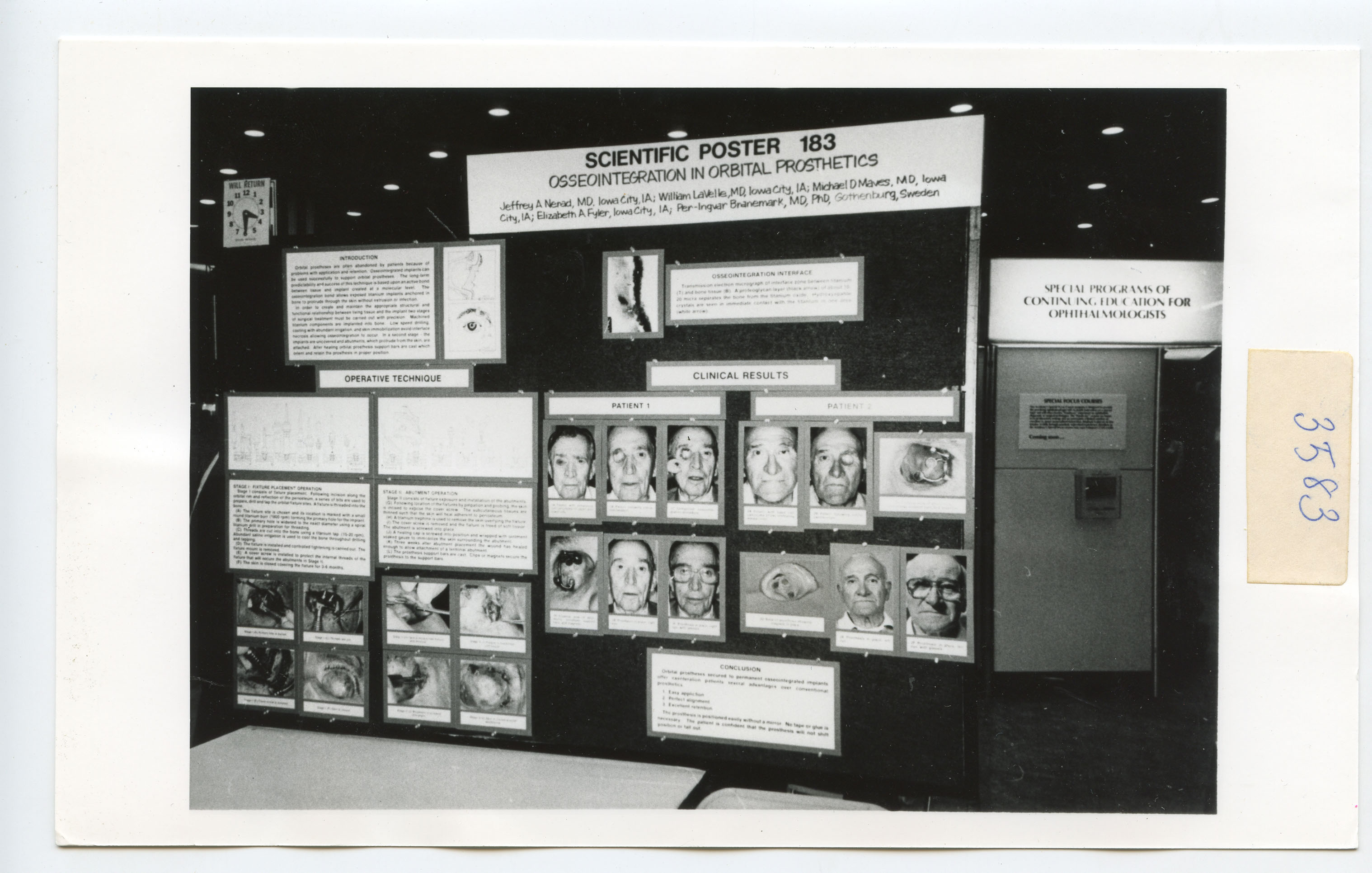
Scientific Poster Presentation, “Osseointegration in Orbital Prosthetics,” Fall 1987, National Museum of Dentistry, ACP Brånemark Collection
Per-Ingvar Brånemark
Early Life and Career
Replacing Teeth
Osseointegration: Full Speed Ahead!
Later Life & Legacy
Acknowledgements & Additional Resources

Per-Ingvar Brånemark: Becoming the Father of Osseointegration has been made possible thanks to the generous gift of the Brånemark archival and object collection by Mrs. Barbro Brånemark to the American College of Prosthodontists and their subsequent donation of the collection and generous support to the museum.
hours
Tuesday - Friday: 10am to 4pm
Address
31 S. Greene St. Baltimore, MD 21201
nmd@umaryland.edu
Phone
410-706-0600
The Dr. Samuel D. Harris National Museum of Dentistry is an auxiliary enterprise of the University of Maryland, School of Dentistry at the University of Maryland, Baltimore.


